4. Boronated Polymers, Liposomes, and Other Nanoparticles as Boron-delivery Systems

Author: Martin Kellert and Evamarie Hey-Hawkins

© Dr. Christoph Selg

Alternative 10B delivery systems including boron-containing polymers , emulsions and liposomes have been investigated as selective boron delivery agents. These approaches involve ligands targeting certain receptors which are overexpressed on the surface of specific tumor cells.

         4.1. Boronated Polymers and Micelles
         4.2. Boronated Biopolymers
         4.3. Emulsions
         4.4. Liposomes
         4.5. Boronated Nanoparticles
         4.6. Boronated Nanotubes

4.1. Boronated Polymers and Micelles

Core cross-linked and carborane-containing micelles (CL micelles) using the radical polymerization of poly(ethylene glycol)-block-poly(lactide) copolymer (PEG-b-PLA) were synthesized (Fig. 4.1a). The results of 125I-labeled CL micelles administrated i.v. into tumor-bearing BALB/c mice suggested suitable accumulation in tumor. Furthermore, tumor growth of mice injected with CL micelles was suppressed for 25 days after neutron irradiation.

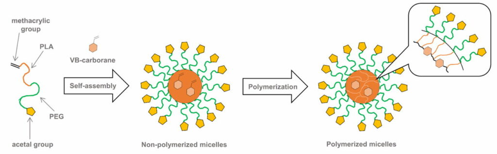
Fig. 4.1a Schematic illustration of the CL micelles containing PEG-b-PLA copolymer

Doxorubicin -loaded carborane-conjugated polymeric nanoparticles (DOX@PLMB) (Fig. 4.1b) were prepared to combine the treatment effects of BNCT and anticancer drug-based chemotherapy. Indeed, the DOX@PLMB nanoparticles selectively delivered boron atoms and doxorubicin to the tumor site of mice simultaneously and enhanced the tumor suppression efficiency compared to doxorubicin-loaded polymeric nanoparticles.

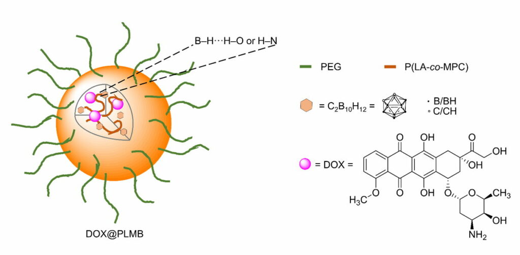
Fig. 4.1b Schematic illustration of DOX@PLMB with its components

Furthermore , three types of size-controlled amphiphilic carborane-conjugated polycarbonate nanoparticles, PN50, PN90, and PN150 (50, 90, and 150 nm in diameter, respectively), were prepared and tested. Sized-dependent cell uptake was observed: smaller size nanoparticles exhibited higher accumulation in tumor cells both in vitro and in vivo, resulting in higher in vivo BNCT effect after neutron irradiation.

4.2. Boronated Biopolymers

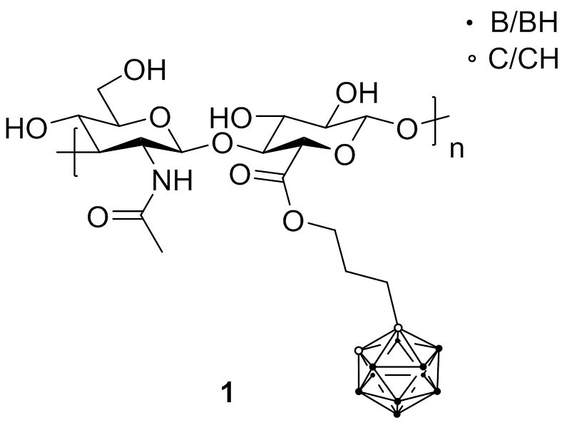
Hyaluronic acid (HA) is a glycosaminoglycan distributed widely throughout connective, epithelial, and neural tissues. As one of the chief components of the extracellular matrix, HA contributes significantly to cell proliferation and migration, and may also be involved in the progression of some malignant tumors. Therefore, HA has emerged as a promising candidate for intracellular delivery of various therapeutic and imaging agents due to its ability to recognize specific cellular receptors, mainly the CD44 antigen that is overexpressed in many cancer histotypes.
First attempts to combine boron sources with HA were conducted with carboranes (1). Biological studies revealed that the boronated HA retained the capacity to specifically interact with CD44 receptor and be internalized to achieve an elevated boron atom content within cells.

Fig. 4.2 HA-carborane conjugate 1

4.3. Emulsions

Lipiodol is an iodinated and esterified lipid of poppy seed oil and is co-administered with chemotherapeutic agents for transcatheter arterial chemoembolization. The intra-arterial administration of an BSH/lipiodol emulsion provided selectively high 10B concentrations (approximately 200 ppm 6 h after administration) in experimental liver tumors. This high 10B accumulation in the liver tumor can be attributed to the embolizing effect of lipiodol on tumor vessels. As an alternative, a water-in-oil–water (WOW) emulsion has also been used as carrier of anti-cancer agents in arterial injections in clinical cancer treatment. For the treatment of hepatocellular carcinoma, a BSH-entrapped WOW emulsion was administered by arterial injections via the proper hepatic artery. The antitumor activity of the emulsion was compared with that of the BSH/lipiodol emulsion or BSH solutions, using VX-2 rabbit hepatic tumor models. The concentration of 10B in VX-2 tumor on delivery of WOW emulsion was superior to that of conventional BSH/lipiodol emulsion. Electron microscopic images confirmed the accumulation of fat droplets of WOW emulsion in the tumor site, but there was no accumulation of fat droplets in the BSH/lipiodol emulsion.

4.4. Liposomes

The development of lipophilic boron compounds embedded within the liposome bilayer is an attractive means to increase the overall incorporation efficiency of boron-containing species, as well as to raise the gross boron content of the liposomes in the formulation. In 1991, BSH encapsulated in a liposome which was conjugated with mAb specific for carcinoembryonic antigen (CEA) was investigated as a liposomal delivery system. This immunoliposome was shown to bind selectively to human pancreatic carcinoma cells (AsPC-1) bearing CEA on their surfaces, and to inhibit tumor cell growth on thermal neutron irradiation in vitro. Furthermore, the cytotoxic effects of locally injected BSH-encapsulated immunoliposomes on AsPC-1 xenografts in nude mice were evaluated. After i.t. injection of the immunoliposomes, boron concentrations in tumor tissue and blood were about 50 ± 6 and about 0.3 ± 0.1 ppm, respectively. Tumor growth in mice that received an i.t. injection of BSH-encapsulated immunoliposomes was suppressed with thermal neutron irradiation in vivo. In 1992, boron-encapsulated liposomes with mean diameters of 70 nm or less from distearoylphosphatidylcholine (DSPC) and cholesterol were prepared. Hydrolytically stable borane anions, B10H102−, B12H11SH2−, B20H17OH4−, B20H193− and B20H182− (two isomers), were encapsulated in the liposomes as soluble sodium salts. The highest boron concentrations in tumor reached the therapeutic range (>15 μg of boron per g of tumor) while maintaining high tumor/blood ratios (>3). The most favorable results were obtained with the two isomers of B20H182−. These boron compounds have the ability to react with intracellular components after they have been deposited in tumor cells by liposomes, thereby preventing the release of the boron compounds into blood. Selective boron delivery to tumors by lipophilic species incorporated in the membranes of unilamellar liposomes was demonstrated by Hawthorne and coworkers in 1995 . They synthesized a nido-carborate amphiphile (K[nido-7-CH3(CH2)15-7,8-C2B9H11], MAC) and prepared boronated liposomes composed of distearoylphosphatidylcholine (DSPC), cholesterol, and MAC in the bilayer. After injecting liposomal suspensions into BALB/c mice bearing EMT6 mammary adenocarcinomas, the time course of the biodistribution of boron was examined. At low injected doses normally used (5–10 mg 10B/kg), a peak tumor boron concentration of 35 μg of boron per g of tumor and a tumor/blood boron ratio of ~8 was achieved. The incorporation of both MAC and the hydrophilic species, TAC (Na3[ae-B20H17NH3]), within the same liposomes significantly enhanced biodistribution characteristics as exemplified by the maximum tumor boron concentration of 50 μg of boron per g of tumor and the tumor/blood boron ratio of 6. This MAC-TAC liposome was administered i.v. in oral cancer model hamsters. After 48 h, the boron concentration in tumors was 67 ppm whereas the precancerous tissue contained 11 ppm. Neutron irradiation resulted in an overall tumor response of 70% after 4 weeks. A high boron content in liposomes was achieved by encapsulating closo-dodecaborate derivatives, Na2[B12H12], Na[B12H11NH3], and BSH. The use of spermidin ium (spd) as a counter cation of closo-dodecaborates was essential not only for the preparation of high boron content liposome solutions but also for efficient boron delivery to tumors. Maximum boron concentrations of 202.7 and 82.4 ppm in tumor were achieved 36 h after injection at doses of 100 and 30 mg[B]/kg, respectively. Furthermore, 100% and 70% survivals were observed in tumor-bearing mice treated with spd-[BSH]-encapsulating liposomes at doses of 30 and 15 mg [B]/kg, respectively, 100 days after BNCT. Transferrin (TF) -receptor-mediated endocytosis is a normal physiological process by which TF delivers iron to cells, and a high concentration of TF receptor has been observed on most tumor cells in comparison with normal cells. Therefore, BSH-encapsulating TF-conjugated PEGylated liposomes (TF-PEG liposomes) were developed and shown to maintain a high 10B level in the tumor, with concentrations exceeding 30 μg of boron per g of tumor for at least 72 h after injection. On the other hand, plasma 10B level decreased, resulting in a tumor/plasma ratio of 6.0 at 72 h after injection. The administration of BSH encapsulated in TF-PEG liposomes at a dose of 5 or 20 mg 10B/kg followed by neutron irradiation suppressed tumor growth and improved long-term survival compared with PEG liposomes, bare liposomes, and free BSH. The biodistribution of BSH- and Na2[B10H10]-encapsulated TF-PEG liposomes in SCC VII tumor-bearing mice was evaluated. The time courses of the change in 10B concentration in the tumors loaded with either of the two liposomes were similar, except that 10B concentrations were higher at 24 h for Na2[B10H10]- than BSH-encapsulated TF-PEG liposomes, and 10B concentration in the tumors was 35.6 μg of boron per g of tumor when Na2[B10H10]-encapsulated TF-PEG liposomes (35 mg 10B/kg) were injected.

4.5. Boronated Nanoparticles

Silver and gold nanoparticles have recently received considerable interest as possible vehicles for drug delivery because of their peculiar physicochemical properties, such as surface plasmon resonance in the visible or near infrared region, photoluminescence , and tunable chemical functionalization through bonding with thiol, amine or carboxylic acid groups.
Water-soluble carborane-functionalized silver nanoparticles conjugated with anti-EGFR antibodies were developed. Surface-enhanced Raman scattering (SERS) microscopy revealed that the modified silver nanoparticles (Fig. 4.5) were selectively targeting an extracellular domain of the EGF receptor. It was estimated that a single 25 nm nanoparticle (surface area ~2000 nm2) with a 45% carborane coating would carry approximately 9000 carborane molecules per targeted antibody.

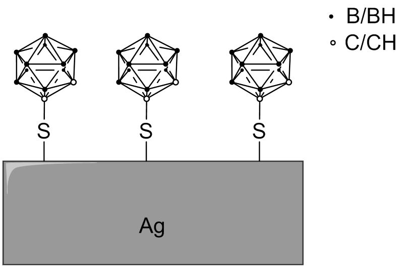
Fig. 4.5 Schematic surface of a silver nanoparticle modified with 1-mercapto-1,2-dicarba-closo-dodecaborane

Furthermore, also gold nanoparticles were applied as boron carriers. For instance, gold nanoparticles, multi-functionalized with BPA, folate, and fluorescent dye for imaging were prepared employing the layer-by-layer coating method. Confocal fluorescence microscopy demonstrated significant uptake of the functionalized gold nanoparticles in folate receptor -overexpressing cancer cells.

4.6. Boronated Nanotubes

As functionalized single-wall carbon nanotubes (SWCNTs) are able to cross cell membranes and concentrate in a number of neoplastic cells without obvious toxic effects, nido-carborates were attached to SWCNTs to produce water-soluble macromolecular boron carriers (Fig. 4.6a). In vivo studies with mice bearing an EMT6 mammary cancer tumor showed 21.5 B/g tumor and a tumor-to-blood ratio of 3.12 : 1.

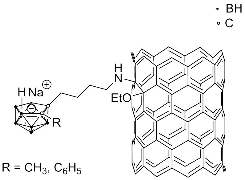
Fig. 4.6a Example of a SWCNT modified with nido-carborate

Boron nitride nanotubes (BNNTs) are analogs of CNTs, in which the carbon atoms are substituted by alternating boron and nitrogen atoms. It was shown that folate-conjugated BNNTs (Fig. 4.6b) exhibited an increased up-take by HeLa cells , probably because of receptor-mediated endocytosis, and co-localized with the lysosomes in the cells.

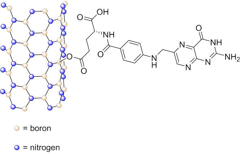
Fig. 4.6b Schematic representation of folic acid conjugated to a BNNT